Getting a brain tumor diagnosis feels like the ground has vanished beneath your feet. One minute you are dealing with headaches or memory lapses; the next, you are staring at an MRI scan that looks like a map of a foreign country. The biggest hurdle isn't just the word "tumor"-it is the alphabet soup of medical jargon that follows. Doctors will mention grades, mutations, histology, and protocols that sound like code.
You do not need to be a neuroscientist to understand your diagnosis. You just need to know what the numbers mean and how they change your treatment plan. This guide breaks down the current standards for classifying brain tumors, explains why molecular testing matters more than ever, and outlines the realistic options available today. We will move past the fear of the unknown and look at the concrete data that guides modern care.
The Shift from Looks to Genetics
For decades, doctors classified brain tumors based on how cells looked under a microscope. If the cells looked messy and aggressive, it was a high-grade tumor. If they looked organized, it was low-grade. That system worked okay, but it missed the engine running under the hood. A tumor could look mild but behave like a wrecking ball, or vice versa.
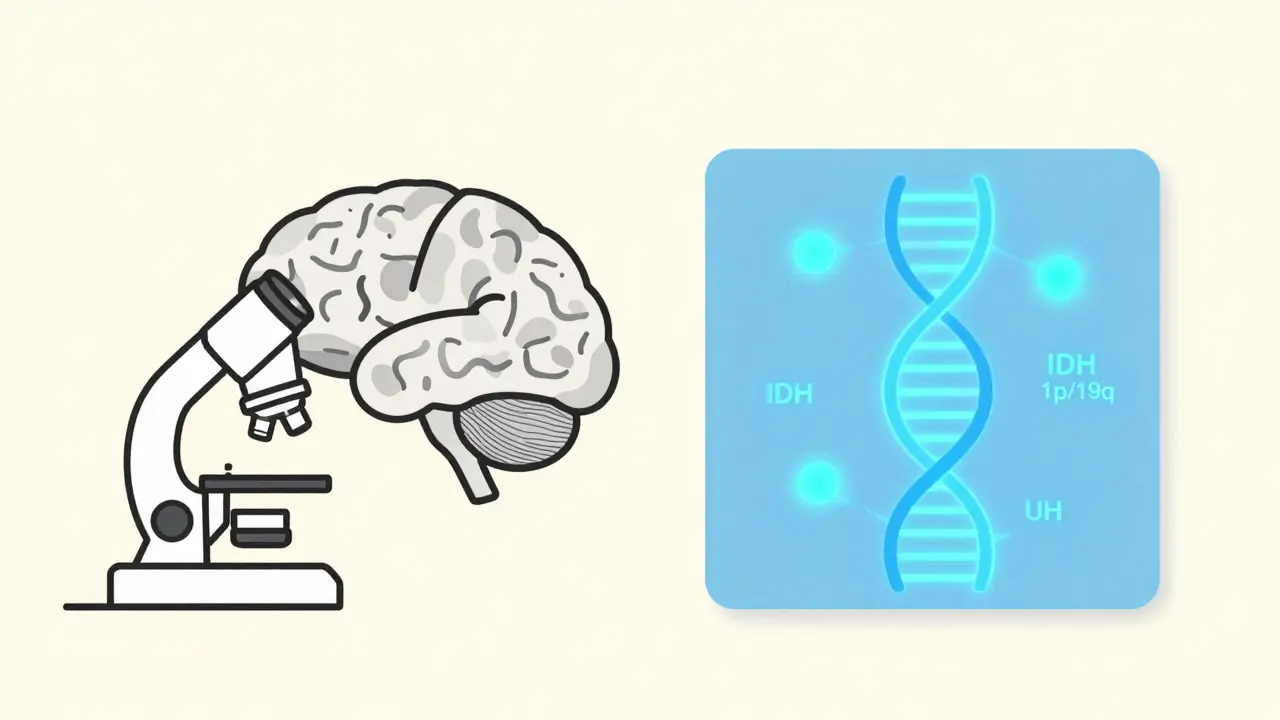
Everything changed with the release of the WHO Classification of Tumors of the Central Nervous System (CNS5) in 2021. This fifth edition marked a massive shift. Instead of relying solely on appearance, the World Health Organization now requires molecular diagnostics to define many tumor types. This means looking at the DNA and RNA of the tumor cells.
Why does this matter to you? Because two patients can have tumors that look identical under a microscope but have completely different outcomes based on their genetic makeup. The new system integrates markers like IDH mutation status and 1p/19q codeletion. These aren't just lab curiosities; they determine whether a drug will work, how fast the tumor might grow, and what your survival statistics look like. Dr. David Schiff from the University of Virginia Cancer Center noted that integrating these markers improved diagnostic accuracy by 35-40% compared to using histology alone. That is a huge leap in precision.
Decoding the Grade: It Is Not Just a Number
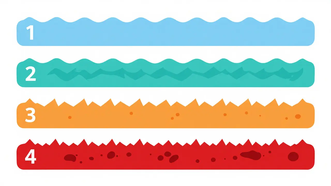
The grade tells you how aggressive the tumor is. The WHO uses Arabic numerals (1 through 4) instead of Roman numerals to reflect that tumor behavior exists on a spectrum, not in rigid boxes. Here is what those numbers actually mean for your life.
| Grade | Growth Speed | Cell Appearance | Treatment Approach |
|---|---|---|---|
| Grade 1 | Very Slow | Nearly normal cells | Surgery often curative; observation may follow |
| Grade 2 | Slow | Slightly abnormal, infiltrating | Surgery + possible radiation/chemo; watch for progression |
| Grade 3 | Moderate/Fast | Abnormal, actively dividing | Surgery + radiation + chemotherapy |
| Grade 4 | Rapid | Highly abnormal, necrotic areas | Aggressive multimodal therapy (Stupp protocol) |
Grades 1 and 2 are considered low-grade. They grow slowly and usually stay contained. However, "slow" does not mean "benign" in the sense of harmless. Grade 2 tumors can infiltrate normal brain tissue, making complete removal difficult. They also carry the risk of transforming into higher-grade tumors over time. For example, a Grade 2 oligodendroglioma might sit quietly for years before becoming a Grade 3.
Grades 3 and 4 are high-grade and malignant. Grade 3 tumors, sometimes called anaplastic, show cells multiplying quickly and invading nearby tissue. Grade 4 is the most aggressive category. These tumors form their own blood supply to fuel growth and often develop dead centers (necrosis). Glioblastomas fall exclusively into Grade 4. They account for more than half of all gliomas and represent the most common cancerous brain tumors in adults.
Major Tumor Types and Their Molecular Signatures
Not all brain tumors are created equal. The type of cell the tumor originates from dictates its behavior. The CNS5 classification groups tumors into specific entities based on both cell type and genetic markers.
Gliomas arise from glial cells, which support neurons. This group includes astrocytomas and oligodendrogliomas. Under the new rules, an astrocytoma must be tested for an IDH mutation. If it has the mutation, it is classified as "astrocytoma, IDH-mutant" and can be Grade 2, 3, or 4. If it lacks the mutation (IDH-wildtype), it is almost always a Grade 4 glioblastoma. This distinction is critical because IDH-mutant tumors generally respond better to treatment and have longer survival rates.
Oligodendrogliomas are rarer and require two specific genetic changes: an IDH mutation AND a 1p/19q codeletion. If both are present, the tumor is an oligodendroglioma, typically Grade 2 or 3. These tumors tend to grow slower than astrocytomas and respond well to chemotherapy, particularly PCV (procarbazine, lomustine, vincristine).
Meningiomas are different. They start in the membranes covering the brain, not the brain tissue itself. Most are benign (Grade 1) and slow-growing. However, about 15-20% are higher grade (2 or 3) and can be invasive. Meningiomas are graded separately from gliomas, ranging from 1 to 3.
The Multimodal Treatment Landscape
Treating a brain tumor rarely involves just one tool. The standard approach is multimodal, combining surgery, radiation, and systemic therapy. The goal is maximal safe resection-removing as much tumor as possible without damaging critical functions like speech or movement.
For Grade 4 glioblastomas, the gold standard remains the Stupp Protocol, established in 2005 and updated in 2017. This involves surgery followed by concurrent radiation and temozolomide (a chemotherapy pill), then maintenance temozolomide for six cycles. While this protocol extends median survival to about 14.6 months, it is grueling. Patients face fatigue, nausea, and immune suppression.
However, the landscape is shifting. In June 2023, the FDA granted accelerated approval to Vorasidenib for adults with IDH-mutant Grade 2 gliomas. This oral targeted therapy blocks the mutated IDH enzyme. The INDIGO clinical trial showed remarkable results: patients taking vorasidenib had a median progression-free survival of 27.7 months compared to 11.1 months for those on placebo. For many, this means delaying radiation and chemotherapy, preserving quality of life for significantly longer.
Other treatments include tumor treating fields (TTFields), which use wearable devices to deliver low-intensity electric fields that disrupt cell division. Clinical trials continue to explore immunotherapies and CAR-T cells, though these remain largely experimental outside of research settings.

Navigating Diagnosis and Costs
Understanding the biology is one thing; navigating the healthcare system is another. Getting a definitive diagnosis takes time. According to the College of American Pathologists, it typically takes 7-10 business days from biopsy to final diagnosis. This delay is due to the complexity of molecular testing.
Comprehensive molecular profiling-including IDH status, 1p/19q codeletion, and MGMT promoter methylation-adds significant cost. Johns Hopkins reports these tests add approximately $3,200 to $5,800 to the bill. While expensive, this investment pays off. Knowing your MGMT status, for instance, predicts how well you will respond to temozolomide. If your MGMT promoter is methylated, the chemo works better. If not, your doctor might consider alternative strategies.
Patient experiences highlight the emotional toll of this process. A 2022 survey by The Brain Tumour Charity found that 68% of UK patients experienced diagnostic delays exceeding eight weeks. Low-grade tumor patients waited an average of 14.2 weeks, while high-grade cases averaged 9.7 weeks. During this wait, uncertainty reigns. Many patients report confusion over grading terminology, with nearly half mistakenly believing a "Grade 2" meant a 20% chance of survival. Clear communication from your care team is essential to bridge this gap.
What Comes Next?
The field of neuro-oncology is moving faster than ever. Liquid biopsies, which detect tumor DNA in cerebrospinal fluid, showed 89% sensitivity in a 2023 study, offering a less invasive way to monitor recurrence. New guidelines from the European Association of Neuro-Oncology (EANO) now incorporate these molecular parameters into routine care.
If you or a loved one faces a brain tumor diagnosis, focus on three things: get the full molecular profile, seek care at a center with high volume experience (pathologists need about 17 cases to master the new WHO system), and ask specific questions about how the grade and type influence treatment choices. Knowledge is not just power here; it is part of the treatment plan.
What is the difference between WHO Grade 2 and Grade 3 brain tumors?
Grade 2 tumors are low-grade, meaning they grow slowly and cells look slightly abnormal. They can infiltrate normal tissue but often allow for long-term survival with monitoring or mild intervention. Grade 3 tumors are high-grade and malignant, with cells dividing rapidly and invading nearby tissue aggressively. Grade 3 tumors typically require immediate, intensive treatment including surgery, radiation, and chemotherapy, and they have a higher likelihood of recurring or progressing to Grade 4.
Why is IDH mutation status important in brain tumor diagnosis?
IDH (isocitrate dehydrogenase) mutation is a key molecular marker used in the WHO CNS5 classification. Its presence helps distinguish between different types of gliomas. For example, an IDH-mutant astrocytoma generally has a better prognosis and responds differently to treatment than an IDH-wildtype glioblastoma. Additionally, the presence of an IDH mutation makes a patient eligible for targeted therapies like vorasidenib, which specifically inhibit the mutated enzyme.
How long does it take to get a brain tumor diagnosis after a biopsy?
According to guidelines from the College of American Pathologists, it typically takes 7 to 10 business days from the time of biopsy to receive a final diagnosis. This timeframe allows pathologists to perform necessary histological examinations and molecular testing, such as determining IDH status and 1p/19q codeletion, which are now required for accurate classification under the WHO CNS5 system.
What is the Stupp Protocol for treating glioblastoma?
The Stupp Protocol is the standard of care for newly diagnosed glioblastoma (Grade 4). It involves three phases: first, maximal safe surgical resection of the tumor; second, concurrent radiotherapy and daily oral temozolomide chemotherapy for six weeks; and third, maintenance cycles of temozolomide for up to six months. This multimodal approach aims to extend survival, with a median survival rate of approximately 14.6 months.
Are there new treatments for Grade 2 gliomas?
Yes, vorasidenib received FDA accelerated approval in June 2023 for adult patients with IDH-mutant Grade 2 gliomas who cannot undergo further surgery. Clinical trials showed it significantly extended progression-free survival to 27.7 months compared to 11.1 months with placebo. This targeted therapy allows some patients to delay more toxic treatments like radiation and traditional chemotherapy.
What does 1p/19q codeletion mean in an oligodendroglioma?
1p/19q codeletion refers to the loss of parts of chromosomes 1 and 19 in the tumor cells. Along with an IDH mutation, this genetic signature is required to diagnose an oligodendroglioma under the WHO CNS5 classification. Tumors with this codeletion tend to be more responsive to chemotherapy, particularly the PCV regimen, and generally have a better prognosis than other types of gliomas.